

J. Mol. Pathol. 2024, 5(2), 215-227; https://doi.org/10.3390/jmp5020014 - 16 May 2024
Abstract
►
Show Figures
The hepatobiliary system is vital for the biotransformation and disposition of endogenous molecules. Any impairment in the normal functioning of the hepatobiliary system leads to a spectrum of hepatobiliary diseases (HBDs), such as liver cirrhosis, fatty liver, biliary dyskinesia, gallbladder cancer, etc. Especially
[...] Read more.
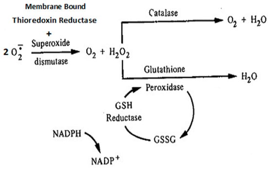
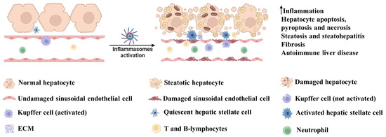
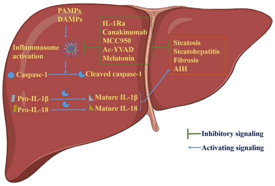
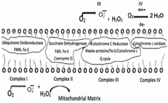
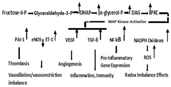
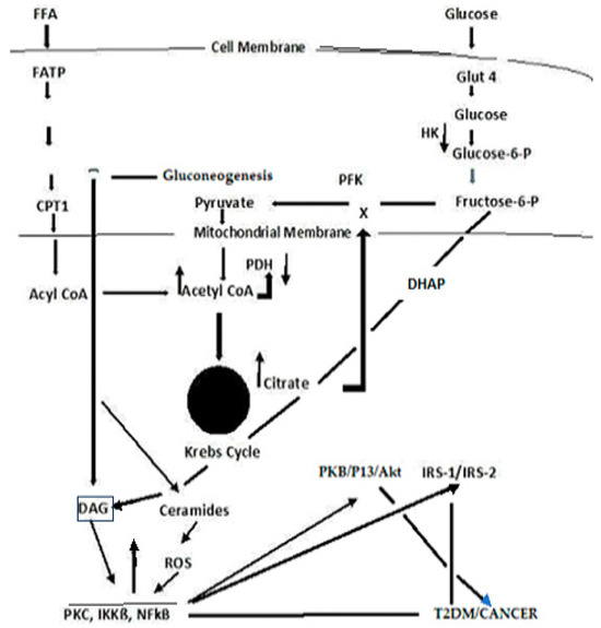
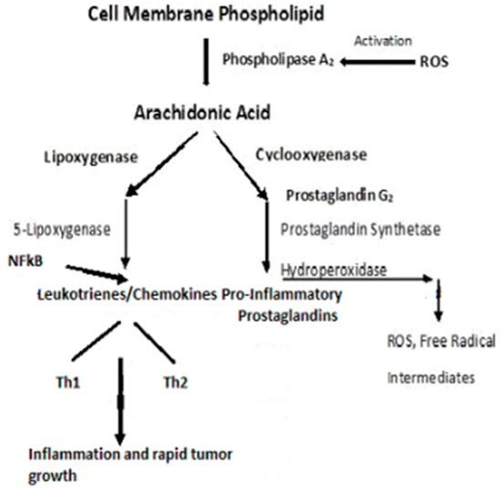
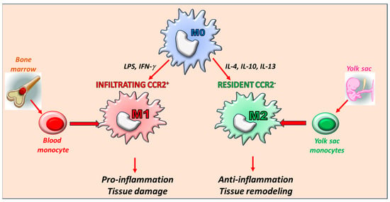
The hepatobiliary system is vital for the biotransformation and disposition of endogenous molecules. Any impairment in the normal functioning of the hepatobiliary system leads to a spectrum of hepatobiliary diseases (HBDs), such as liver cirrhosis, fatty liver, biliary dyskinesia, gallbladder cancer, etc. Especially in pregnancy, HBD may result in increased maternal and fetal morbidity and mortality. Maternal HBD is a burden to the fetus’s growth, complicates fetal development, and risks the mother’s life. In fetal programming, the maternal mechanism is significantly disturbed by multiple factors (especially diet) that influence the development of the fetus and increase the frequency of metabolic diseases later in life. Additionally, maternal under-nutrition or over-nutrition (especially in high-fat, high-carbohydrate, or protein-rich diets) lead to dysregulation in gut hormones (CCK, GLP-1, etc.), microbiota metabolite production (SCFA, LPS, TMA, etc.), neurotransmitters (POMC, NPY, etc.), and hepatobiliary signaling (insulin resistance, TNF-a, SREBPs, etc.), which significantly impact fetal programming. Recently, biotherapeutics have provided a new horizon for treating HBD during fetal programming to save the lives of the mother and fetus. This review focuses on how maternal impaired hepatobiliary metabolic signaling leads to disease transmission to the fetus mediated through the gut–brain axis.
Full article

Figure 1


